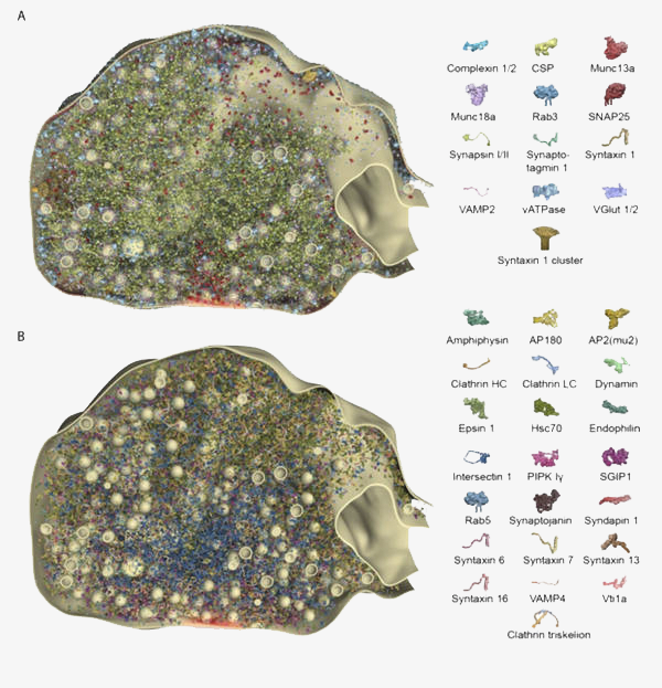
The organization of exo- and endocytotic proteins within a model synaptic bouton. Sections through the synaptic bouton in which only the exocytotic (A) or the endocytotic proteins (B) are shown. The graphical legend indicates the different proteins (right). The graphical legend indicates the different proteins (bottom). Displayed synaptic vesicles have a diameter of 42 nm.
About
In the first funding period we analysed the dynamic partitioning of proteins in the presynaptic bouton. Among other results, we found that soluble protein mobility was strongly affected by interactions with the synaptic vesicle cluster. This induced a clear separation between the protein composition of the synapse and that of the rest of the axon, and thus acted as the gate, or composition-proofing structure, of this system. In the next funding period we will analyse the postsynaptic compartment, focusing on the glutamatergic dendritic spine. We will test two hypotheses: 1) The 3D structure of the spine, including the neck, acts as a diffusion barrier that participates in retaining molecules, thus forming a morphological gate; 2) The PSD and/or spine organelles influence the localization of soluble proteins, forming a proteinaceous gate. We will use several complementary approaches for this, from live imaging to super-resolution imaging and computer modelling.


